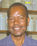
 Cell biologist George M. Langford is the Ernest Everett Just Professor of Biology at Dartmouth College. He grew up in rural North Carolina and received a B.S. degree from Fayetteville State University. He earned a Ph. D. from the Illinois Institute of Technology and did postdoctoral training at the University of Pennsylvania. Before arriving at Dartmouth, Dr. Langford held positions at the University of Massachusetts at Boston and at the medical schools of Howard University and the University of North Carolina. He has served as program director for the cell biology program of the National Science Foundation (NSF) and is a member of the National Science Board. He has made important discoveries about how organelles move inside cells; he also works to combat the under-representation of minorities in science. We met at the Marine Biological Laboratory (MBL) in Woods Hole, Massachusetts, where Dr. Langford spends his summers.
How did you choose science as a career?
It was actually by a process of elimination. I've always loved the arts, especially music, but in high school, I realized that I just didn't have the training to pursue a career in music. Luckily, I became interested in science and had some wonderful teachers who nurtured my interest. My high school didn't have good science facilities—North Carolina was still segregated. But perhaps because there weren't a lot of opportunities for blacks, many talented people became teachers. And the science teachers tried to encourage promising students. When I got to college, I decided to major in biology.
What drew you to cell biology?
In high school math, I had been particularly good at geometry, and it was shapes and structures that first attracted me to cell biology. Even as an undergraduate I loved microscopy, and I was good at visualizing the three-dimensional structures represented by two-dimensional images. This was the era when the electron microscope was first revealing the fine details of cell structure.
Who were some of your mentors during your graduate and postgraduate training?
I was fortunate to be able to do my Ph. D. research under Bill Danforth, who ran one of the strongest biology research groups at my university and was a supportive mentor. My research was on the biochemistry of glucose metabolism in Euglena—not microscopy. But while I was there, I was a teaching assistant for a famous embryologist, Jean Clark Dan. She urged me to do postdoctoral research with Shinya Inoue, a reknowned microscopist at Penn. Shinya taught me the importance of choosing the best experimental system for the question being asked. At his suggestion, I studied the movement of an exotic organelle, the axostyle, that certain protozoa use for swimming. It's made of microtubules, tiny protein tubes just like the ones in cilia [see figure 1.12]. But unlike cilia, the axostyle is entirely inside the cell and causes the entire protozoan surface to undulate.
You went on to be a medical school professor. What led you to go to Dartmouth?
Dartmouth had just established a professorship in honor of Ernest Everett Just, a black alumnus who had graduated in 1907. I was invited by Dartmouth to apply for that position, and I discovered that I liked the community there very much. Also, I missed undergraduate teaching. So I left Chapel Hill for the cold north.
Who was Ernest Everett Just?
He was a developmental biologist and a pioneer who worked hard to break down barriers for black scientists in this country. His mother, a school teacher, had managed to enroll him in a prep school that sent most of its graduates to Dartmouth. Just was a stellar student there and at Dartmouth. After graduating and earning a Ph. D. at the University of Chicago, he did important research on fertilization and early development in animals, working mainly with marine invertebrates. At the time, genetics was an exciting new field in biology, and many biologists thought that embryonic development was completely controlled by the nucleus. It was Just who established the crucial role of the cytoplasm in fertilization and the early events of development. He showed that there were important signals going from the cytoplasm to the nucleus, as well as the other way around.
Did he achieve recognition in his day?
No, he experienced racial discrimination that eventually led him to emigrate to Europe; he returned to the United States only when the Nazis took over Germany. He was very dispirited, and he became ill and died at 59. I think his heart was broken by his failure to get a position at a major American research university. His graduate training had been with F.R. Lillie—for whom this MBL building is named—and Lillie was a father figure for him. But when he asked Lillie to recommend him for a position at a white university, Lillie was unwilling. By the way, in the summer Just worked with Lillie right here at the MBL.
What exactly is the MBL?
The MBL is an independent research institution owned and governed by the scientists who work here. Founded in 1888, it has had an enormous impact on biology in America, because so many biologists have come here to do research or take a course. It's a major gathering place for biologists in the summer, and there are now year-round programs, too.
Is research limited to marine organisms?
No, lots of scientists here work on mice or other nonmarine animals. But a number of us do come here to work with marine organisms. I use the nervous system of the squid in my research, primarily because of the squid's giant axon.
What is a giant axon, and how do you use it in your research?
An axon is a long, thin extension of a nerve cell that carries electrical signals from the cell body (the vicinity of the nucleus) to another cell, which can be far away. The speed of electrical conduction along an axon is proportional to its thickness. Squids benefit from this principle by having an unusually thick axon that connects their brain area to muscles in their mantle, which they use for locomotion. When the squid sees a predator, it can signal its muscles very quickly and escape. The so-called giant axon results from the fusion of about a thousand ordinary axons. The length of the giant axon we use is about 25 centimeters.
The question we're investigating is the mechanism by which molecular "motors" move cargo around in the cell, using protein filaments as tracks. The filaments I'm concerned with are microtubules (just like the ones in cilia and axostyles) and thinner filaments called actin filaments, which are two of the elements of a structural network called the cytoskeleton. Molecular motors are proteins that utilize chemical energy from ATP to do mechanical work—such as moving an object along a track.
Now let's return to nerve cells and their axons. The cell body of a nerve cell produces a lot of materials that need to be transported to the end of the axon. For example, neurotransmitter molecules are synthesized and packaged in vesicles (small sacs) for eventual release at synapses [see figure 2.18]. Molecular motors are responsible for transporting these vesicles from the cell body to the axon terminals, and they do so using filaments of the cytoskeleton as tracks. We use the squid giant axon for studying this transport not only because of its size but also because you can strip away the plasma membrane and leave the cytoplasm intact with transport still going on. We can then watch and record the filaments and vesicles by high-resolution video microscopy, which cannot be done with other cells. And we can easily test the effects of different chemicals on transport.
We also use the methods of molecular biology, including recombinant DNA technology. For instance, we've cloned the gene for the motor protein we're working on, called myosin-5, and we can use bacteria to produce the different parts of the motor separately. We can then add different combinations of these parts to our giant-axon preparations to determine what the various parts do.
What's the most exciting discovery you've made using squid giant axons?
When we first started looking at vesicle transport in giant axons, it was thought that the only filaments involved were microtubules. But one day, as we were observing vesicles moving under the microscope, we suddenly noticed that there were vesicles moving in areas of the preparation lacking microtubules. And we said, aha, something interesting is going on here—there have to be other kinds of filaments supporting the transport, ones not visible with our microscope. Actin filaments were a prime candidate. To make them visible, we labeled them with a fluorescent dye. Sure enough, fluorescing images of actin filaments then showed up in the "empty" regions where the vesicle transport was taking place.
Based on those observations, we proposed a dual transport mechanism, by which transport vesicles use microtubules as expressways and actin filaments as local streets. The vesicles move down the axon along microtubules, but once at the axon terminal, they transfer onto actin filaments to get to the plasma membrane.
What questions are you addressing now?
We're trying to learn how a vesicle transfers from a microtubule to an actin filament. We know that there are two different kinds of motors: The motor molecule that moves a vesicle along a microtubule will not work on an actin filament. So there has to be a switch from one motor to the other. We've learned that the two motors interact directly with each other, and we're now trying to understand this interaction.
Do you also study other kinds of cells?
I'm using mammalian kidney and lung cells in collaborating with a group at Dartmouth that studies cystic fibrosis (CF), a devastating genetic disease. The CF defect is in a protein called CFTR, which normally is located in the plasma membrane and helps transport chloride ions across. We're studying how CFTR gets from its site of synthesis to the membrane, a process that requires molecular motors. In most CF patients, the CFTR protein has a defect that prevents it from reaching the membrane. If we can figure out exactly how CFTR is transported, we may be able to develop a treatment to help patients.
Tell us about some of your efforts on behalf of minorities in science.
A legacy of the racism encountered by Ernest Just continues to discourage minorities. While at the NSF, I attacked this problem at the postdoc level by starting a program to help minority Ph. D.'s get the advanced training and mentoring they need to develop research careers. Later, at Dartmouth, I set up the E. E. Just Program, with the goal of raising the number of minority science majors. We have a forum that brings students together with professors, who talk about what excites them in science and serve as resources and mentors for the students. We also provide internships enabling students to do research in faculty labs. We're now seeing an increase in the number of minority science majors, though there is still more to be done.
©2005 Pearson Education, Inc., publishing as Benjamin Cummings