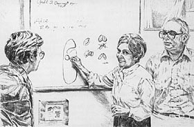
Interview: Peter and Birgit Satir
The discoveries of Birgit and Peter Satir are elegant examples at the cellular level of a fundamental theme of this unit and of biology in general: Structure fits function. Birgit is interested in how cells secrete chemical products, and she has been able to correlate this function with specialized regions (microdomains) of cell membranes. For most of his career, Peter has studied cilia and flagella, which function in cell motility. The cilia and flagella of all cells except bacteria share a common architecture consisting of structures called microtubules arranged as a circle of nine pairs surrounding two single microtubules, in what is known as the 9 + 2 arrangement. Peter has described how the sliding of microtubules past one another causes the undulations of cilia and flagella.
Both Peter and Birgit Satir are professors of cell biology in the Department of Anatomy and Structural Biology at Albert Einstein College of Medicine. In one segment of the following interview the Satirs discuss how they, as spouses trained in the same academic field, overcame the difficulties of being accepted as independent researchers.
Birgit, at what point in your education did you decide on a career in biology?
BS: Growing up in Denmark, I attended something called Gymnasium, where students were exposed at the age of 18 to various fields. During a lecture in a big auditorium, a physiologist demonstrated nervous reflexes in the leg of a frog whose head had been cut off. I was absolutely fascinated; I wanted to understand how it all happened. That was the impetus for me to change to biochemistry from a language major. The following summer, I took courses in math, physics, and chemistry to catch up to students who had majored in science. I went on to get a Ph.D. in biochemistry at the University of Copenhagen. At the time, biochemistry was a new major, and there were only two other students besides myself. We had to design our own program of courses.
So it was through biochemistry that you began to study cells?
BS: I was doing such things as isolating RNA, but for me it was not enough to study isolated molecules. I wasn't satisfied just looking at artificial membranes; I wanted to know how things happened in a cell. I wanted to know how the molecules of the cell interact and what the molecules do in the cell.
Peter, how did your interest in biology evolve?
PS: Well, my early experience was very different from Birgit's. When I started at the Bronx High School of Science in 1949, biology was the first science course I took. The first day of class, my biology teacher had us look at Paramecium with the microscope. When you look at Paramecium, the first thing you see is that it moves. And it moves with its cilia. I remember asking my teacher, "How do the cilia move?" He said, "Nobody really knows how they move." At the age of 14, I had asked the question that still dominates my research interests. Of course, I had a long way to go to be a biologist.
What was your route?
PS: I went from Bronx Science to Columbia University, where I was the only zoology major. Most of the other students taking life science courses were pre-med. Now, we're talking about the year 1954, the year of the Watson-Crick model of DNA, and I got caught up in the excitement the model was creating at Columbia and the whole excitement of biology. I took a course called Protozoa as Living Systems, the only time the course was ever given. It was really my first experience in the laboratory, and I saw all those beasts [paramecia] running around again. I renewed my interest in cilia. During my senior year, I took a seminar course in cell motility and chose ciliary movement as my seminar topic. I had decided I wanted to go to graduate school, which was unusual at that time.
In the 1950s, before Sputnik, the country produced only about a hundred Ph.D.'s per year. Rockefeller Institute, a research institute in New York City, had just started its graduate program and asked the president of Columbia to nominate a student. When I was invited to apply to the institute, I knew exactly which laboratory I wanted to join. While reading the literature in preparation for my seminar on ciliary movement, I found that the major current articles were published by Keith Porter, a cell biologist at the Rockefeller Institute who was using an electron microscope to study the ultrastructure of cilia and other organelles of the cell. I would finally have an opportunity to do research on a question that had intrigued me since high school.
So you and Birgit were graduate students at the same time, but on opposite sides of the Atlantic. How did you meet?
PS: The president of Rockefeller Institute believed that science was an international occupation that transcended national boundaries. Graduate students were encouraged to spend a year abroad. I went to Eric Zeuthen's laboratory at the Carlsberg Biological Institute in Denmark. Birgit was Zeuthen's graduate student at that time.
BS: The Carlsberg Biological Institute had become a little international research center. We had a stream of foreign visitors. It was a very lively place, with many seminars and discussions. I was working on cell division for my thesis when Peter joined our lab. We thought he was incredibly spoiled. We still had economic problems from the Second World War. When Peter came as a graduate student, his income from his stipend was about the same as the head of the laboratory, who had three children. Peter was even able to buy a car. Peter went back to the United States before we got married. I wanted to stay in Copenhagen to finish my degree. I got my Ph.D. in 1961, the same year Peter finished his graduate work at Rockefeller.
PS: That summer, Birgit and I both attended an international conference in Stockholm, where we got engaged. Birgit joined me in New York the next year, and we moved to Chicago together. I had accepted a position as an instructor at the University of Chicago. Those were the days of very strong nepotism rules. At the University of Chicago, husband and wife were allowed to work in the same department, but not under the same supervisor.
BS: And at that time, women were usually shuttled off into positions as research associates and rarely got regular faculty positions that could lead to tenure. I had a half-time position as a research associate. It was a difficult period of adjustment for me, but I managed to survive.
PS: We moved to Berkeley in 1967. We were the first couple in the history of the University of California at Berkeley to be allowed to work in the same department.
BS: Once again, however, I was limited to a research associate position with little independence. I had essentially no chance of getting a faculty appointment. For seven years as a research associate, I was not allowed to apply for my own research grant through the university. I was finally able to establish my independence by going outside the university to apply for a special fellowship. Obtaining this outside support also entitled me to apply for my own research funding from federal agencies. That's what really started my career.
Here at Albert Einstein College of Medicine, you are both professors, tenured faculty members, in the same department. Did the opportunity for these separate but equal appointments help to attract you to Einstein?
BS and PS: Absolutely!
PS: The growing women's movement has made a big difference at many universities. But Albert Einstein has a long tradition of recognizing and promoting women scientists and of offering individual faculty positions to men and women who happen to be spouses. It also has a tradition of husband-and-wife teams.
BS: There's an important difference here. A team is two people who work on the same thing. So we're not really a husband-and-wife team because we don't do that; we have independent academic careers.
To return to the earlier part of your career, what were you working on when you were at Berkeley?
BS: Inspired partly by Peter's work, my interest was shifting to cell ultrastructure, the detailed structure of the cell as revealed by the electron microscope. And I began to get interested in membranes, because the freeze-fracture techniques for studying them had just been developed. I was especially interested in the structure and function of the membranes of the cell. That led to my work on exocytosis, the process by which many cells secrete chemical products. For example, cells in the human pancreas secrete digestive enzymes by exocytosis. Before the products are secreted, they are stored within the cell in vesicles bounded by membranes. During exocytosis, the membrane of a secretory vesicle fuses with the plasma membrane, the membrane at the surface of the cell, and the substance within the vesicle is deposited outside the cell. At Berkeley I became interested in the question of how these secretory vesicles fuse with the plasma membrane.
PS: It was a very exciting time at Berkeley. Daniel Branton had done the groundwork on the freeze-fracture method, which was a new way of preparing cells for examination with the electron microscope. The technique fractures a cell membrane along its middle, separating the two molecular layers of the membrane and revealing the membrane's structural organization. Birgit learned the freeze-fracture method and made her discovery.
BS: I combined freeze-fracture images with the ultrastructure and then stimulated the cell to secrete to see if the membranes reflected the changes that must occur during membrane fusion. The regulation, the control mechanism, is what I'm still working on. One thing I found earlier is that the plasma membrane has specialized sites where secretory vesicles fuse during exocytosis. These sites are marked in the membrane by a ring of about nine to eleven protein particles surrounding another particle. We call these sites "fusion rosettes," and they are located just above secretory vesicles that will fuse with the plasma membrane. The particles of the rosette disperse in the membrane when the vesicle fuses. It was fascinating to discover that the membrane has microdomains, local regions specialized in structure and function.
Birgit, the cells you have used in most of your studies of exocytosis are ciliates, unicellular organisms such as Paramecium and Tetrahymena. It is often perplexing to an outsider why a research biologist chooses a particular organism to study a certain process. Why are ciliates well suited to the investigation of secretion by exocytosis?
BS: Beneath the plasma membrane of one of these single-celled organisms are rows of secretory vesicles. When the organism encounters a threat, the cell discharges the contents of all of these vesicles en masse. The exocytosis can be experimentally induced. It is an excellent system for studying the mechanism of exocytosis and how the process is controlled. Also, there are several different genetic mutations in these ciliates that block secretion at one step or another. This provides an opportunity to dissect the secretion apparatus. By comparing how these various mutations affect the structure of the fusion rosette and the process of exocytosis, we hope to determine the key steps regulating secretion by cells. Although I am studying secretion in ciliates, because they are ideal experimental organisms for the questions I am asking, I believe that the mechanism of exocytosis is similar for other cells, including human cells, that secrete products.
PS: I chose to study the clam gill, which turns out to have been the classical system for studies of ciliary motility. It was largely a practical decision. If you wanted to know how mammalian cilia work, you could look at the trachea. But it's hard to get the tissue. And you're involved with animals, which means that you have to worry about which animals, animal care, the treatment of animals. Nobody worries about how you open your clams!
In addition, with mammalian systems, you have to worry much more about temperature control, carbon dioxide control, and oxygen control than you do with, say, a ciliate or invertebrate organs. And when you look at a mammalian trachea, you can hardly see the cilia. They're so small and they're covered with a blanket of mucus. You see something move, but you don't know what's going on. When you look at a clam gill, you see cilia. So you're already miles ahead. That's why the clam gill was chosen in the first place and why I stuck with it.
It is the faith of cell biologists that although cells differ in overall structure, the component parts of cells, the organelles, work much the same in one cell as they do in another.
That faith has been justified by your own work on the structure and function of cilia, Peter.
PS: Well, the universal ultrastructure of cilia and flagella in all eukaryotic organisms was one of the things that attracted me to these organelles. Don Fawcett and [Keith R.] Porter, my mentor, discovered the ordered array of microtubules within cilia that became known as the 9 + 2 arrangement.
I was fascinated by how this organelle had been conserved in essentially the same form, whether located in Paramecium or other ciliates; the gills of clams or other invertebrates; or the windpipe, oviduct, or sperm of humans. The construction must be a very useful one to have persisted without substantial change since its evolutionary inception. I concentrated on correlating the structure of the cilium with how the locomotory organelle works.
I think one of the difficulties students have when they first begin to study cells is comprehending how microscopic structures can contain subdivisions such as organelles with different functions or microdomains. Spending so much time exploring the microscopic world of cells, you must become accustomed to a scale of sizes not too meaningful to most people. Are there any tricks you can share that may help students to develop some sense for the microdimensions of cells and their parts?
BS: This is always a problem for students. At the other extreme is the problem of trying to comprehend vast distances, such as the distance to a star. In dealing with either very small or very large dimensions, you must have frames of reference on which to base comparisons of size. One standard we use in studying the ultrastructure of cells is the cell membrane, which has a thickness of about a hundredth of a micrometer.
PS: There's another aspect of this problem of scale. In studying cells, we have to continually remind ourselves that we are working in a different dimension, where ordinary concepts don't necessarily apply. Our common sense about how things work in the macroscopic world around us cannot always be extrapolated to the cell.
I'll use the function of cilia in cell motility as an example. Someone once published a paper in which he said cilia move the way a human being does the breaststroke in a swimming pool. Well, fluid dynamics are very different in the microscopic realm. To a cilium, water is about as viscous as liquid asphalt would be to a swimmer. Textbooks often describe cilia as working like little oars or paddles. But a ciliated cell actually crawls through water, using its cilia to push the water. So cilia are very good for moving mucus, which is a very viscous substance. And in the realm we normally deal with, if you stop swimming or rowing, you glide for a while. On the other hand, when its cilia stop beating, the cell doesn't glide, but stops right on the spot because of the viscosity of the environment. This is the reason microorganisms can respond instantaneously compared to the world we know.
Let's shift downscale still another notch. Each of you, Birgit and Peter, has published papers on the molecular basis of the processes you study in cells. Do you anticipate that recent breakthroughs in molecular biology, such as recombinant DNA technology, will have any impact on your research?
PS: I think the distinction between cell and molecular biology is becoming more and more blurred. Ciliary movement and exocytosis both involve specific interactions between various protein molecules, which of course are gene products. We would like to know more about the synthesis, structure, function, and evolution of these proteins. Birgit is further along than I in thinking of ways to apply molecular biology to her research.
BS: I just spent a sabbatical at California Institute of Technology learning new methods in molecular biology that may help me study the roles of certain proteins in exocytosis, particularly in the regulation of membrane fusion. In my work, recombinant DNA technology will be very useful for reading out the structure of specific proteins and for designing changes in the protein structure for new experiments. For as long as I have focused on exocytosis in my research, there have always been new challenges, new questions to ask, and new methods that make those questions approachable.
PS: In my own case, I have stuck with one cellular organelle, the cilium, throughout my career. This is not so narrow as it may seem. The study of cilia has implications for biology at the level of whole organisms, at the level of ciliated tissues, and at the cellular and molecular level. Just now, for example, an exciting area is the movement of membrane-bound vesicles along microtubules in nerve axons, which seem to involve force-producing mechanisms related to those in cilia. I believe that by standing at one point in biology and asking appropriate questions, you can have an impact on problems concerning the many levels of biological organization.
©2005 Pearson Education, Inc., publishing as Benjamin Cummings