Interview: Charles Leblond

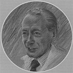
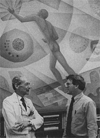
Over the mantel in the century-old anatomy library at McGill University in Montreal is an imposing portrait of Professor Charles Leblond, one of Canada's most famous scientists. Dr. Leblond was born in France in 1910 and received his M.D. degree there before moving to Canada, where he joined the anatomy department at McGill during the early 1940s. One of the pioneers of modern cell biology and histology (the study of tissues), Professor Leblond has spent most of his life studying the dynamic renewal of the body's cells and their subcellular components. In this interview, Dr. Leblond traces the historical development of this work.
The 1950s and '60s, when electron microscopes and other new instruments became available, were golden decades in the study of cell structure and function. What was it like to be involved in cell biology during that exciting period?
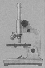
Well, my own interest was in using radioactive isotopes to localize key compounds in cells and tissues. I mostly did this work at the level of the simpler light microscopes. But at the time, we were always thinking, "Ah, if we could only go a little farther, if we could only see more details in the cells." And when we saw the first pictures coming out of Keith Porter's lab—beautiful pictures taken with the electron microscope showing all these details within cells—that was really fantastic.
How can we help our students rescale their spatial thinking to the microscopic level, so they can better understand the cell and its parts?
This is a very important problem in teaching biology. In our anatomy and histology courses here at the medical school, we try to take the students through changes in scale in a series of steps: for example, from the stomach wall, which you can see with the naked eye, to the tissue layers and cells of the wall, which you can see with the light microscope, to the organelles within the cells, which you can see only with the electron microscope. This gradation makes the scale changes less extreme. We never show the students pictures taken with the electron microscope without also showing them photographs taken with the light microscope for orientation. It's difficult, but eventually students adjust to the different scales at which we study life.
You mentioned that you helped develop techniques for localizing chemical compounds within cells and tissues. How is this done?
The technique is called radioautography or, more commonly, autoradiography. Radioactive atoms or molecules are introduced into organisms or cells via injection. Most radioisotopes behave in the body in the same way as nonradioactive isotopes of the same chemical elements, except researchers can detect the presence of the radioisotopes, of course. So cells use their normal metabolism to process these radioactive tracers. For example, cells take up radioactively labeled amino acids and incorporate these precursors into proteins. These labeled proteins can then be localized within tissues and cells by placing thin sections of the cells on a glass slide and coating the sections with photographic emulsion. The sections are then placed in the dark for a certain period, and the radiation emanating from radioactive sites in the cells exposes the photographic emulsion. The slide is developed and this preparation, now called a radioautograph, is examined with the microscope. Black silver grains from the photographic emulsion are seen directly over the sites in the cell containing the products made from the labeled precursors. So radioisotopes function in research as spies that report what is going on in the body.

You helped develop this technique?
Well, the first radioautographs were made by Antoine Lacassagne in Paris in the 1920s. He injected radioactive polonium into rabbits, embedded the organs in paraffin, and then placed these paraffin blocks in a dark room with photographic plates kept in contact with the surfaces of the blocks. He used small weights to keep pressure on the plates. He found that the polonium was mostly concentrated in lymph nodes, but the results were very crude. Lacassagne invited me to join his laboratory in Paris in the 1930s, and I began my work on the behavior of iodine in the body.
Using radioautography?
Yes. Well, first, I actually used a Geiger counter to locate radioactive iodine that had been injected into animals. This demonstrated that the iodine was concentrated in the thyroid gland. Then I tried Lacassagne's radioautography, except, instead of using the whole organ in a paraffin block, I cut sections of the block and placed the sections of thyroid on a glass slide. Then I put a photographic plate on the slide. I was able to show that the tracer had been incorporated into the colloidal material within the follicles of the thyroid gland. But the resolution was still poor.
How did you improve the technique?
A few years later, after I was discharged from military service in 1946, I returned to McGill University. A histologist named Leonard BeLanger and I then concentrated on improving radioautographs. Instead of using a photographic plate, we found that we could melt the emulsion off film and then paint this emulsion directly onto our radioactively labeled sections of tissues. The improvement was dramatic, and we began to localize tracers in tissues at high magnification with the light microscope. Later, of course, it was possible to examine radioautographs with the electron microscope and study the passage of radioactive labels through the different organelles of cells.
Can you give some examples?

Well, we were able to show how the cells of the thyroid gland produce, store, and secrete the thyroid hormone, which regulates metabolism in the body. In other experiments, we used radioautography to study the metabolic functions of the organelle called the Golgi apparatus. By using chemical precursors labeled with radioisotopes, we learned that the Golgi is where the cell adds sugars to proteins. This turns out to be one of the most important functions of the Golgi. There were many other discoveries. My lab at McGill was like a beehive: so many researchers using these new techniques that we didn't even have time to publish all our results, although we've managed to publish well over 300 papers since that time.
In one of your articles, you wrote that the use of radioisotopes has added a time dimension to cell biology. Can you explain?
Before radioactive isotopes were first used in this century to trace the chemical dynamics of cells, the prevailing view, especially among anatomists, was that the human body was composed of organs that were stable, with chemical constituents that were permanent for as long as the person lived. There seemed to be some support for this view. For example, in the late 1800s, several German scientists showed that the amount of nitrogen an organism ingests as food protein is equal to the amount of nitrogen leaving the body in feces and urine. The conclusion was that the organs kept their own proteins intact, and simply broke down the proteins from food and got rid of the nitrogenous waste products. The body, in this view, was like a locomotive that simply consumed fuel in the form of food. The organs were like the cogs and wheels of the locomotive, using the food to do work, but not actually incorporating any of the molecules of the food into their own structure. The use of detectable isotopes to trace chemicals in the body overthrew this old view of a stable body with organs that were static in their chemical makeup.
How?
In the late 1930s, Schoenheimer injected animals with amino acids labeled with a heavy isotope of nitrogen. Less than a third of this nitrogen subsequently turned up in the feces and urine. The other two-thirds of the injected radioactive nitrogen was incorporated into the organs of the body. About the same time, Georg de Hevesy, who won a Nobel Prize, injected rats with radioactive phosphate and found that this labeled phosphate was incorporated into bone tissue and also turned up in phospholipids and other chemical components of various tissues. These experiments demonstrated the dynamic turnover of substances making up cells, but did not provide precise locations for the movement of these transient chemicals through the cells, tissues, and organs. That's where radioautography came in and introduced the time dimension into cell biology.
So some of our organs are constantly changing their cellular parts?
Yes. For example, we were able to show that all of the cells of the stomach lining are replaced every few days. Yet, even as the new cells replace the dead cells that are sloughed from the lining, the histological pattern, the arrangement of cells in the tissue of the stomach lining, is unchanged. Six hundred years ago, Thomas Aquinas realized that the components of the human body are always changing (with age, for instance), but he also pointed out that the identity of the individual is retained. The same can be said of the dynamic states of cells and tissues: There is constant turnover of components, but the same cell types and arrangement of cells in the tissue are continuously regenerated. Methods that give cell biology a time dimension are critical to our understanding of this renewal process.
You mentioned how you began your research in cell biology in France soon after finishing medical college. When did you come to Canada?
Well, before Canada, I went to Yale on a Rockefeller fellowship, where I continued my work on cell biology. I also got involved in a very different type of project. On weekends, I went down to New York, to the American Museum of Natural History, where I became quite friendly with the head of the amphibian section. This was during the depression, and he had 23 WPA workers helping him. I helped find things to keep them busy. Twenty-three people, some of them Ph.D.'s, and there they were, out of work. Some of them were German immigrants, and so I put them to work translating Konrad Lorenz's book on animal behavior. That was how Lorenz's great work first became widely known in America. Lorenz later received the Nobel Prize.
It was also during my period in the United States that I met and married my wife, Gertrude, who is American. Then the war started, and I was in the French Army. For complicated reasons, I was a private, even though I was a medical doctor. I was sent to Morocco, but I managed to get out after six months at the end of 1940. It was then that McGill University offered me a job in their anatomy department. And at the same time, I signed up with the Free French Army. So just a few years after I began working in Montreal, the Free French sent me to London to collaborate with British scientists on some research involving psychology and the selection of personnel for the war effort. My only connection to psychology was that I had once done some experiments on how injection of certain hormones induced maternal behavior in rats. That didn't seem to have much to do with personnel selection, but at least I was able to keep intellectually active during the war. After the war, I received a telegram from McGill asking me to come back and I've been here since 1946.

The bulk of your life's work has been accomplished in Canadian universities. Are they very different from their counterparts in the United States?
Not anymore. Over the past century Canadian universities have changed gradually from the British system to an American system. When I arrived at McGill before the war, students still addressed their professors as "sir." By the end of the war, that formality had disappeared.
How do undergraduates prepare for medical education in Canada?
Now that's a little different here in Quebec compared to the United States and even the rest of Canada. After high school, all college students spend their first two years at a junior college, taking general education courses. From there, students can go on to a university and complete their four-year degree. But many premedical students spend only one year in university studies before entering medical school. During that year, they mostly study chemistry, physics, and biology. In the United States, I think, students graduate from college before entering medicine. About half of our students do the same, which I think has advantages. Students are more mature with another year of undergraduate studies.
After you finished medical school in 1934, why did you decide to concentrate on basic research rather than medicine?
Actually, I wanted to do research before I went to medical school. But my family wouldn't hear of it when I told them I wanted to be a scientist. Then I said, "Well, maybe I'll go into medicine." They were delighted. They thought that by the time I finished medical school, I would forget about science. My small medical school in France wanted me to stay there after graduation and eventually join the medical faculty. But I still had no intentions of practicing medicine. I did practice for about three months. I needed some money. That type of work just didn't appeal to me as much as research did. You see one patient, and then another, and then another, and at the end of a good day of hard work you have seen 25 patients. And then the next day you see 25 other patients. Many doctors like the variety. Myself, I'd like to work with one patient for several days. In any case, I never had any doubt about wanting to be a scientist and having the fun of doing research.
©2005 Pearson Education, Inc., publishing as Benjamin Cummings