Interview: Shinya Inoué
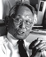
Part biologist, part engineer, Shinya Inoué has helped reinvent the use of light microscopes in the study of cells. Building his own microscopes, Professor Inoué invented optical technology that reveals the structure and movement of minute organelles without having to kill and stain cells. For example, such technical virtuosity enabled Dr. Inoué to confirm the existence of structures called spindle fibers, which separate chromosomes during cell division. In 1992, the American Society of Cell Biology recognized Dr. Inoué's contributions with its highest honor, the E. B. Wilson Award. Professor Inoué was elected to the prestigious National Academy of Sciences in 1993. He presently holds the title of Distinguished Scientist at the Marine Biological Laboratory (MBL) in Woods Hole, Massachusetts. Each summer, scientists from around the world converge at MBL in order to share ideas and collaborate on their research. Science is social, and the Woods Hole culture has helped shape modern biology. At least 34 Nobel laureates have been part of the MBL community. In this interview, Shinya Inoué shares some of his thoughts about MBL and about the microscopic world of cells.
What is the Marine Biological Laboratory, and how did it start?
It started in 1888 as an offshoot of the Women's Educational Association of Boston. U.S. fisheries researchers first recognized that the coast off Woods Hole is a place where the warm current and the cold current mix, and therefore it is very rich in life. MBL came shortly after. It was started essentially as a summer research station with a collection of scientists from different universities. Scientists came here not just to study marine life per se, but to use marine organisms as appropriate models for studying various basic aspects of life.
Why do you think the lab has been such a magnet for top scientists who seem to migrate here each summer?
The ambiance certainly contributes. But a tradition has built up over the years of people coming together and exchanging ideas. All the lectures are open to everybody in the community. The summer courses are taught by top-notch scientists. And we have a world-famous library.
Team research seems to be common at the MBL.
Well, none of us are experts in enough fields. Collaborations are extremely important. I think in order to have a fruitful collaboration, you need to have people who know not only their own field but enough of another field to have a feeling for it. Biologists, physicists, and chemists collaborate naturally at the MBL, where they are free from university chores.
Can you tell us a little bit more about your own career? What attracted you to science?
My father was in the foreign service, so I grew up in different countries. I was born in England, and then we went to Japan briefly, then China, the United States, Australia. From my high school years on, I lived in Japan. Science was one of the things that spanned the different countries—it provided cultural continuity.
Technology and science interested me a lot when I was very young. Biology as such didn't interest me very much in high school, except for one class where the teacher showed us how birds' feathers stayed as neat as they are with simple barbs. The rest was so rote, and memorizing things is not my forte. There was so much memorization in the Japanese school system. In college, which is between high school and university in Japan, I had very good science teachers who were researchers, and one, Professor Katsuma Dan, really sparked my interest. He challenged us with interesting projects in the biology lab, so it was no longer memorizing things or repeating what others had done, but finding out how you discover things. I think that is when I really got interested in science.
As a cell biologist, you have engineered many improvements in light microscopes. What are the advantages of light microscopes compared to electron microscopes?
About the time I started studying cells, a lot of people went into electron microscopy because of the very high resolution. Certainly we learned a lot from electron microscopes, but in electron microscopy you have to kill, fix (preserve), and slice the cell. You get a still image of a part of a cell, which may or may not be modified during the preparation. With light microscopes, on the other hand, you have a limited resolution but you can see the dynamics of living cells.
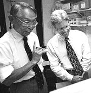
With so many types of light microscopes available from optical companies, why do you build your own?
None of them did the job that we needed to get done. The polarizing microscope does an excellent job of identifying the optical characteristics of crystals, but these microscopes as they came wouldn't work in the small areas of living cells, so I started building. For example, this special objective lens increases sensitivity for detecting where molecules are lined up in the cell.
 When did you build your first microscope?
The first one was while I was still in Japan after the second world war. Emperor Hirohito came visiting to Dan's laboratory at the Misaki Marine Station. Before the war, the Emperor had published volumes of biology, but he had to publish under somebody else's name because the Emperor was considered to be a god. After the war, he got released from being a god and became an ordinary person and a biologist. Anyhow, when he came I was told, "Why don't you show the Emperor some of the specimens swimming around?" I had different bits and pieces of microscopes that were stacked on books to align their axes, but it was too unstable. So I tied them together on a sturdy base and got started on the first version of my microscopes.
And most recently you have been a pioneer in video microscopy.
With video microscopy you can use the light microscope the way it was supposed to work but never did because the contrast became too low at high resolution and shallow depth of field. When you put that image through video and a computer, out pops all kinds of images that weren't possible, such as molecular filaments that are much thinner than the microscope's resolution limit. Another advantage of video microscopy is the time dimension. With video you can have immediate playback of what you have been recording, so you can plan the experiments based on what the recording shows.
Your inventiveness with light microscopy dates back to your studies in the 1950s of spindle fibers and their role in cell division. Tell us a little bit about that work.
In those studies I was able to see the arrangement of molecules inside living cells. By being able to see regions where the molecules were lined up, I could show that chromosomes were moved by fibers attached to them. In textbooks, you see these spindle fibers attached to chromosomes and it looks convincing. But people who were studying mitosis seriously at that time said, "You see those fibers when you fix a cell and stain it, but that doesn't mean they are there in the living cell." In fact if you fix the cell very carefully, you don't see any fibers. There was a 50-year battle raging back and forth as to whether chromosomes were moved by those fibers or had some other means of moving. I was able to show, by improving the polarizing microscope, that these were real structures. I was also able to show why there was such a controversy. Essentially, those fibers are very unstable; if you say "boo" to a cell, they disappear.
How do you think these spindle fibers work—how do the chromosomes move apart?
We don't have a definitive answer yet. The fibers that I saw were bundles of structures called microtubules. I showed that the molecules making up the microtubles were assembling and disassembling. Furthermore, I said that the chromosomes move as the microtubules fall apart. Scientists said, "Well, that's crazy! You can't have a string that is both falling apart and pulling something." I showed these fibers and their behavior in 1950 or 1951, but it took about 15 years before the microtubules could be isolated from cells. When they were isolated, the micro-tubules showed none of the characteristics I predicted they would. It took another 10 years or so until somebody figured out why the isolated fibers would not behave the way the live fibers behave. It turns out that these fibers are terribly sensitive to calcium levels. When you grind up the cell, a huge amount of calcium leaks from stored compartments and destroys the microtubules. A young researcher figured out that you have to sequester calcium that leaks out so it can't attack the spindle. Then he was able to isolate the spindles and show that the molecules do assemble and disassemble. Now, disassembling and shortening microtubules can be made to pull chromosomes even outside the cell.
But there was also very good work showing that there are molecular motors that move on top of microtubules in cilia and flagella. When that came into vogue, everybody wanted to say that chromosomes move because there are molecular motors on the chromosomes that make them crawl along microtubules. How to combine these two different concepts or observations is still up in the air.
What are some of the other important questions about how cells divide?
By using extracts from frog eggs and clam eggs, scientists have been able to show where mitosis can be interrupted. They also have identified chemical compounds produced by the cells that control stages of cell division, for example, when the cell goes into mitosis, when the nuclear envelope breaks down and cell division takes place, and so on. That obviously has an immediate significance in cancer research because in cancer cells, cell division goes wild. Why do some cells go wild and not others? What controls the division?
How does your research on cell division connect to other questions in basic cell biology?
Well, if we look at this example of the microtubules, it turns out that the concept of the microtubules assembling and disassembling applies not just to cell division but also to nerve growth, white blood cells moving around and capturing bacteria, and transport of organelles in cells. All of these things are related. Once we start understanding the basics of how the cell and its components behave, then we have a better chance of understanding the kind of things that many scientists are asking questions about but are having difficulty finding answers to.
What advice do you have for students as they begin to think about the structure and function of cells?
I continue to worry about science being learned as a collection of facts and theories. One needs to have a certain body of knowledge—but in addition, one needs to understand how the knowledge is acquired—that really is at the heart of science.
©2005 Pearson Education, Inc., publishing as Benjamin Cummings